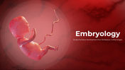
Free - Embryology Template For PowerPoint And Google Slides
Free

 Canva
Canva
Embryology Presentation Template
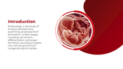
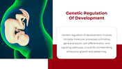
Visualizing how a single cell becomes a functioning human body is the hardest part of teaching anatomy. Traditional textbooks often fail to show the fluid transition between stages. This embryology slide deck provides a logical, step-by-step path through prenatal life, making it perfect for medical lectures, biology classes, or clinical briefings.
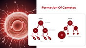
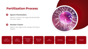
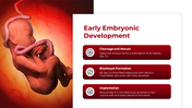
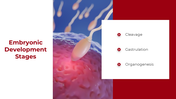
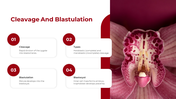
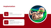
The journey starts at the very beginning: the cellular split of sperm and egg. You can track exactly how 46 chromosomes become 23 before fertilization occurs. From there, the focus shifts to the first critical week. It maps out how a zygote divides, forms a blastocyst, and finally finds its home in the uterine wall to begin a pregnancy.
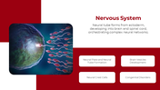
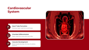
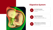
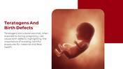
As development continues, the focus moves into specialized organ systems. You can explain the folding of the brain's neural tube, the looping of the primitive heart, and the complex rotation of the gut. To keep the talk grounded in medicine, there is a dedicated look at the amnion and yolk sac. A vital section on birth defects and harmful toxins ensures that the science is always linked to real-world health risks. Every 3D image is built to stay clear even in a large lecture hall.
Download this template to give a fast, accurate, and system-by-system talk on human development.
Features of this template:
- Fully editable slides.
- Works in PowerPoint, Google Slides, and Canva.
- Available in 16:9 and 4:3 formats.
- Chromosome Reduction Maps: Clear views of the 46-to-23 cell split for both parents.
- Primitive Tube Focus: Individual slides for the early nervous, heart, and gut systems.
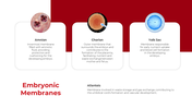
- Fetal Support Layers: Detailed icons for the amnion, chorion, and yolk sac.
- Clinical Health Risks: A specific slide to discuss how teratogens affect a fetus.
- 3D Medical Renders: High-quality images that show exact fetal anatomy and position.
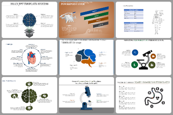
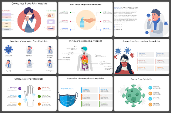
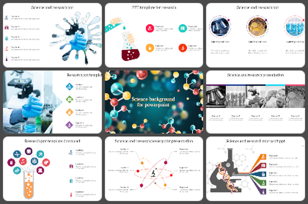
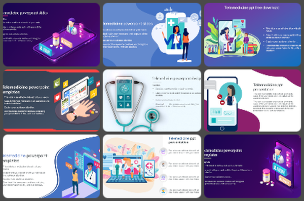
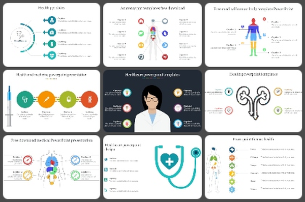
You May Also Like These PowerPoint Templates
Add to Wishlist
Download
Edit

Add to Wishlist
Download
Edit

Add to Wishlist
Download
Edit

Add to Wishlist
Download
Edit

Add to Wishlist
Download
Edit

Add to Wishlist
Download
Edit

Add to Wishlist
Download
Edit

Add to Wishlist
Download
Edit

Add to Wishlist
Download
Edit

Add to Wishlist
Download
Edit


Add to Wishlist
Download
Edit

Add to Wishlist
Download
Edit

Add to Wishlist
Download
Edit

Add to Wishlist
Download
Edit

Add to Wishlist
Download
Edit


Add to Wishlist
Download
Edit

Add to Wishlist
Download
Edit


Add to Wishlist
Download
Edit

Add to Wishlist
Download
Edit

Add to Wishlist
Download
Edit


Add to Wishlist
Download
Edit

Add to Wishlist
Download
Edit

Add to Wishlist
Download
Edit

Add to Wishlist
Download
Edit

Add to Wishlist
Download
Edit

Add to Wishlist
Download
Edit

Add to Wishlist
Download
Edit

Add to Wishlist
Download
Edit


Add to Wishlist
Download
Edit
