Diabetic Retinopathy Template For PPT And Google Slides

 Canva
Canva
Diabetic Retinopathy Presentation Template
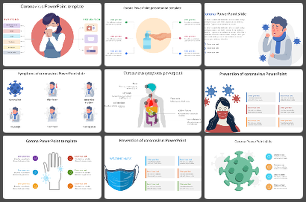
Explaining how high blood sugar damages the eye requires a clear link between microscopic changes and visible symptoms. This presentation deck serves as a technical bridge for ophthalmologists, diabetes educators, and medical students. It focuses on the biological progression of retinal damage, moving from healthy blood vessel anatomy to advanced proliferative stages.
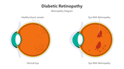
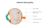
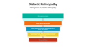
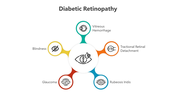
This deck is structured to support a deep clinical narrative. It begins with side-by-side anatomical diagrams comparing a normal eye to one with retinopathy, highlighting hemorrhages and "cotton wool" spots. A core strength of this deck is the pathogenesis funnel, which maps the transition from microvascular occlusion to neovascularization. This allows presenters to explain why the disease progresses, rather than just showing the result.
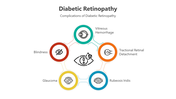
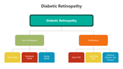
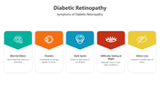
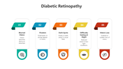
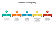
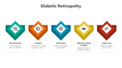
For patient-facing education, this deck includes a comprehensive symptom matrix. You can walk through common warning signs—such as floaters, dark spots, and night blindness—using clean, icon-based layouts. This template also addresses long-term risks, including glaucoma and retinal detachment. By categorizing the disease into Non-Proliferative (NPDR) and Proliferative (PDR) stages, these slides provide a professional framework for discussing diagnosis, risk levels, and the urgency of treatment.
Use this deck to deliver an authoritative, step-by-step briefing on diabetic eye disease and patient risk management.
Features of this template:
- Fully editable slides.
- Works in PowerPoint, Google Slides, and Canva.
- Available in 16:9 and 4:3 formats.
- Retinal Pathology Diagrams: Detailed views of aneurysms, hemorrhages, and hard exudates.
- Stage-Wise Classification: A full breakdown of Mild, Moderate, and Severe NPDR vs. PDR.
- Pathogenesis Flowchart: A five-step visual guide to disease development (Hypoxia to Neovascularization).
- Complication Star-Map: Connects retinopathy to blindness, glaucoma, and vitreous hemorrhage.
- High-Contrast Symptom Icons: Large, easy-to-read markers for vision loss and distorted sight.
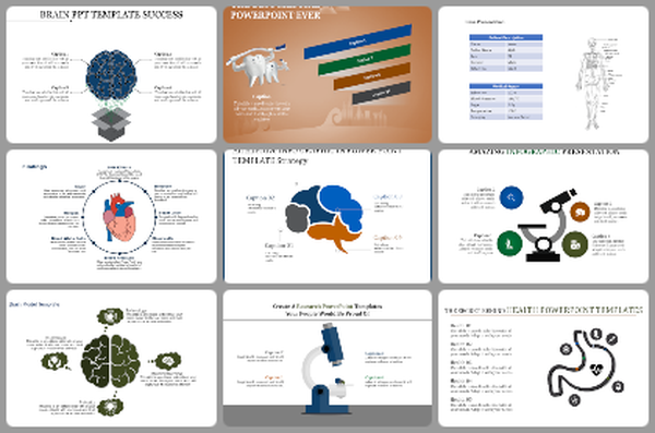


You May Also Like These PowerPoint Templates

Add to Wishlist
Download
AI Customize

Add to Wishlist
Download
AI Customize

Add to Wishlist
Download
AI Customize

Add to Wishlist
Download
AI Customize

Add to Wishlist
Download
AI Customize

Add to Wishlist
Download
AI Customize

Add to Wishlist
Download
AI Customize

Add to Wishlist
Download
AI Customize

Add to Wishlist
Download
AI Customize

Add to Wishlist
Download
AI Customize

Add to Wishlist
Download
AI Customize

Add to Wishlist
Download
AI Customize

Add to Wishlist
Download
AI Customize

Add to Wishlist
Download
AI Customize

Add to Wishlist
Download
AI Customize

Add to Wishlist
Download
AI Customize

Add to Wishlist
Download
AI Customize

Add to Wishlist
Download
AI Customize

Add to Wishlist
Download
AI Customize

Add to Wishlist
Download
AI Customize

Add to Wishlist
Download
AI Customize

Add to Wishlist
Download
AI Customize

Add to Wishlist
Download
AI Customize

Add to Wishlist
Download
AI Customize

Add to Wishlist
Download
AI Customize

Add to Wishlist
Download
AI Customize

Add to Wishlist
Download
AI Customize


Add to Wishlist
Download
AI Customize

Add to Wishlist
Download
AI Customize

Add to Wishlist
Download
AI Customize

Add to Wishlist
Download
AI Customize

Add to Wishlist
Download
AI Customize

Add to Wishlist
Download
AI Customize

Add to Wishlist
Download
AI Customize
